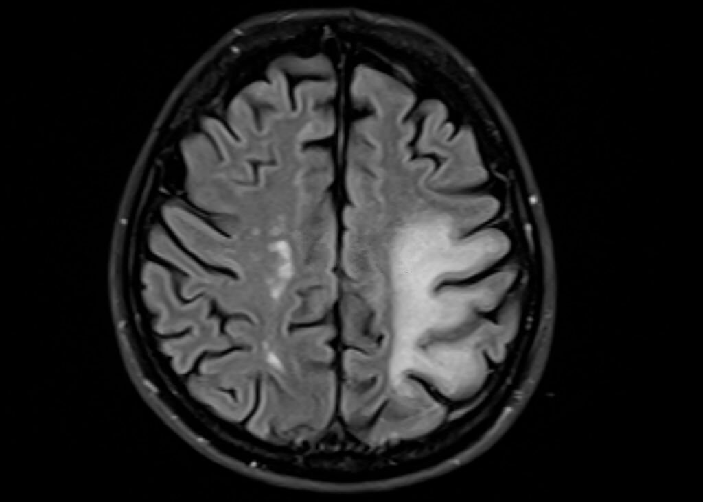
Scientists discovered that reduced “integrity” in the brain stem area was connected to a quicker deterioration in remembering and reasoning in older persons and certain neural alterations identified in earlier Alzheimer’s disease, utilizing several brain scanning technology.
According to the latest analysis, alterations in a section of the brain stem apparent on scanners could be a possible early sign of Alzheimer’s illness.
Early Alzheimer’s Signs In The Brain Stem
Among the top diseases, Alzheimer’s is a leading one, and hence it has been a major point of survey and study by experts also. The brain stem, if properly diagnosed, can help to know this health condition at an early stage, and precautions for the same can be taken immediately.

Genetic markers were substances that could be tested to accurately identify a disease, for example, a molecule in the bloodstream or a result on a medical examination. The study, which was reported in the journal Science Translational Medicine on Sept 22, is the latest in a long-running search for “biomarkers” that can aid in early Alzheimer’s identification.
Scientists were attempting to comprehend the fundamental progression of the illness fully and, in the meantime, develop markers that can detect Alzheimer’s illness earlier, she said. According to Rebecca Edelmayer, managing partner of scientific outreach for the Alzheimer’s Association, “most patients with Alzheimer’s are diagnosed based on evaluations of their memory, reasoning, and other thinking skills.”
Here are several possible methods for doing so, such as brain scans and monitoring particular compounds in the cerebrospinal or bloodstream. Some of these technologies have been researched and utilized in inpatient care.
“Very interesting,” said Edelmayer, who’s not engaged in the recent study.
She believes it identifies a possible early indication that can assist in identifying “normal” brain aging from a pathological one.
But, as Jacobs, an associate professor at Harvard Medical School and Massachusetts General Hospital, stated, it’s unknown if this is a component of an illness. Despite amyloid formation, which occurs later on in life, tau deposition occurs young age. According to Heidi Jacobs, the report’s principal investigator, data reveals that around half of 30- and 40-year-olds had tau buildup in the LC.
The findings are predicated on 174 older persons who are mainly mentally sound. MRI scans were performed on all of them to determine the “integrity” of an LC. Due to its tiny size, Jacobs said, it’s impossible to quantify tau in the LC directly. However, recent developments in MRI technology have enabled a measurement of the area’s coherence, indicating the presence of tau accumulation.
Subjects were also subjected to PET imaging in addition to the MRI examinations. The goal is to look for tau and amyloid buildup in other parts of the brain linked to the earliest stages of Alzheimer’s disease. Eventually, their recall and other cognitive functions are assessed during eight years.
As per Jacobs, this will not establish that tau accumulation in the LC initiates the full procedure. However, it highlights LC integrity as just a possible marker for Alzheimer’s disease progression. Overall, lower LC coherence is linked to tau deposition in the entorhinal cortex, a memory-related brain area. In addition, lower LC coherence is connected to a faster deterioration in research members’ cognitive abilities.
Although most individuals are familiar with the amyloid that characterize Alzheimer’s disease, Edelmayer claims that tau development is most directly linked to memory loss. It’s also possible that a connection among the protein as well as other variables is at play.
“There is a cascade of events that happens 10 to 20 years before the clinical signs of Alzheimer’s,” Edelmayer said.
“Any technology that can reliably detect changes along that path could potentially lead to earlier diagnosis,” she said.