
Keeping life going is the responsibility of the human heart. There are four chambers in the heart. Approximately the size of a clenched fist, the heart is quite large. It is the most complex working muscle in the human body and functions throughout a person’s life.
Hearts Are Forged With Guts
Almost all animals, including humans, have a heart that pumps blood throughout the body. The heart-like pumping organs of invertebrates such as grasshoppers function similarly to that of humans.
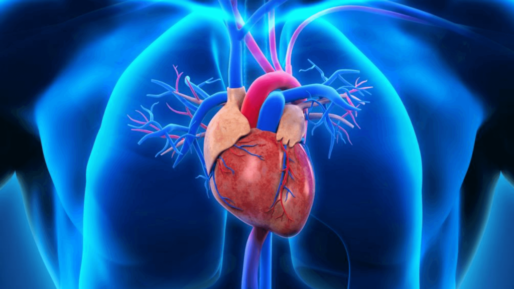
The human heart consists of two ventricles and two atria and is about the size of a human fist. Blood is pumped through the ventricles and received through the atrium. In addition to the right atria and ventricles, the left atria and ventricles make up the left heart. The aorta is the largest artery of the body in the heart.
The deoxygenated blood returns to the heart from the superior and inferior vena cava. Blood is pumped into the right ventricle from the right atrium through the tricuspid valve. The pulmonary semilunar valve is responsible for pumping blood from the right ventricle to the pulmonary trunk.
Its main function is to transport blood to the lungs, release carbon dioxide and absorb oxygen. The pulmonary veins carry blood from the lungs to the heart, and the left atrium is where blood enters the heart from the pulmonary veins.
A muscle wall called the septum separates the right and left sides of the heart. Blood is pumped from the right ventricle to the lungs through pulmonary arteries for reoxygenation.
Blood cannot flow back into the heart through the right semilunar valve since it closes. By way of the pulmonary veins, oxygenated blood is then delivered into the left atrium.
All living things are built from cells. There are trillions of cells in the human body. Each cell is responsible for providing structure and function, absorbing nutrients from food, converting them into energy, and performing specific functions. A cell can also make copies of itself and contain hereditary material.
Cells that appear identical to a newborn begin dividing into distinct types that eventually become a variety of tissues and organs. By exchanging chemical signals, neighboring tissues activate different development steps in each other. Yet, understanding the effects of this crosstalk is complicated.
We developed a brand-new way to grow three-dimensional organoids organ-like clusters of cells, which mimic how the gut and heart tissues cooperatively develop from stem cells.
A new organoid of this type could help researchers understand how tissue signaling influences healthy human development in the laboratory.
In a new study published in the journal Cell Stem Cell, Gladstone Senior Investigator Todd McDevitt, Ph.D., explains that its findings suggest new avenues for understanding how developing organs cooperate and instruct one another.
An Organoid Discovered by Coincidence
McDevitt’s lab has developed methods for growing organoids, which are multicellular tissues that can reproduce in a dish the properties, function, and structure of the heart, brain, lungs, and other organs.
Human-induced pluripotent stem cells are used to create his team’s heart organoids. Various cocktails of nutrients and other naturally occurring substances are used to coax these cells into becoming heart cells. A different cocktail could allow the scientists to create a greater variety of heart cells than the conventional cocktail, which yields mostly heart muscle cells.
They weren’t attempting to create a new type of organoid in their original plan, and they were surprised to find that the new cocktail produced organoids containing both gut and heart cells in several cases.
Ana Silva, Ph.D., a postdoctoral fellow and first author of the study, says they were intrigued by the fact that organoids usually develop into one type of tissue. As with the embryonic stage, heart and gut tissues developed simultaneously.
In the past, researchers have grown single-tissue organoids separately, then attempted to combine them later to examine crosstalk between developing tissues.